Professor Shi Fudong reviewed the report presented at the 2020 Annual Meeting of the National Society of Psychiatry
(September 19, 2020; More than 18,000 neurologists listened to the report online. Authors: Zheng Pei, Chen Jingshan, PhD)
In 2014, the classic science fiction film Super Body was released. If you remember, it tells the story of a professor who researches and develops the potential of the brain. Dolphins use 20 percent of their brain neurons and are born with a sonar system. Humans now use only 10% of the neurons in their brains, and are already so creative that their inventions can fly to the sky and the moon and the sun go together. If we can mobilize 20 percent of our neurons to connect to our limitless potential, we may be able to overcome current crises, diseases, shortages caused by excessive use of energy resources, and global warming that threatens humanity and the homeland of all living things……
Neuroscientists and neurologists are forever curious about the infinite potential of the brain, so how can we have a window to observe the mysterious workings of the brain and re-understand the structure and changes of brain physiology and pathology? Only in this way can we truly understand brain diseases, lay the foundation for effective and targeted treatment, and unleash the limitless potential of medicine for the benefit of patients. Through the recent work of his team, Professor Shifu Dong presented a vivid picture of how neurologists can use classic and most advanced biotechnology as a unique "window" to study neurological diseases at the National Neurological Academic Year meeting on September 19. The magical journey of "Window to the brain" begins……
Figure 1: Windows to the Brain, first page of Professor Shi's report
The first bright light of the journey: Brain aging and immunity
By comparing and screening the immune imprints of brain tissue of young and old people, it was found that the immune cells represented by Natural Killer cells gradually increased in human brain with age. Most of these cells are located in the dentate gyrus region of neurogenesis in the brain and are adjacent to the neural precursor cells (Figure 2). Combined with single cell sequencing and proteomic techniques, this research group further found that some of the neural progenitors of senescence overexpressed various senescence related phenotypes, including interleukin-27. These immune factors can activate and amplify the accumulation of immune cells in the brain by gene coding and pedigree tracing. In addition, RNA sequencing and immune screening and other evidence show that aging leads to downregulation of major histocompatibility complex I molecules (MHC-I) on the surface of neural precursor cells, resulting in immune tolerance loss, thus activating immune surveillance and damaging neural precursor cells during normal brain aging. The elimination of immune cells in the aging brain by immune intervention can promote the survival of neural precursor cells and improve cognitive function. The results will be published in Nature Neuroscience in November 2020. In this study, the relationship between immunity and brain aging was revealed using classical methods, i.e. normal brain tissue combined with new high-throughput methods.
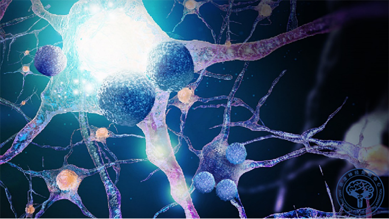
Figure 2: Immune cells and neural precursors in the aging brain. In the neurogenetic region of the aging brain, a loss of immune tolerance causes nerve precursor cells (purple cells with protuberance, pictured) to be cleared by immune cells represented by NK cells (blue globular cells).
"I do not know the true face of Lushan Mountain, only because I am in the mountain."
Acute brain injury causes rapid changes in multiple systems and organs around the body (Liu Qiang, Jin Weina, et al., Immunity 2017). These distant changes can reflect brain damage and thus open a new window on the understanding of brain disease. By analyzing bone marrow cells from patients with cerebral hemorrhage who underwent decompression with bone flap, the team found that the hemorrhage rapidly activated bone marrow hematopoietic stem cells. Combined with micro PET imaging, gene coding, pedigree tracing and other technologies, it was further found that after cerebral hemorrhage, the body generated mononuclear cells with immunosuppression function in a short period of time through innervation. Through Nanostring screening and immunoimprinting, the team also identified molecular switches that mediate the formation of monocytes from hematopoietic cells within the bone marrow. As an autoprotective mechanism after intracerebral hemorrhage, these newborn monocytes migrate to intracerebral lesions and eventually inhibit neuroinflammation and perihematoma cerebral edema. In addition, the use of fDA-approved selective beta3-adrenergic agonists can promote the growth of more immunosuppressive monocytes in the bone marrow, thereby reducing neuroinflammation and improving the prognosis of intracerebral hemorrhage. By identifying changes in distant tissue and cell composition after brain injury, the study reveals that damaged brains can, over the course of evolution, dispatch peripheral cells to the brain for rescue. Neurologists can reduce brain damage by amplifying this mechanism of self-protection (Figure 3). The work will be published in Science Translational Medicine at the end of 2020.
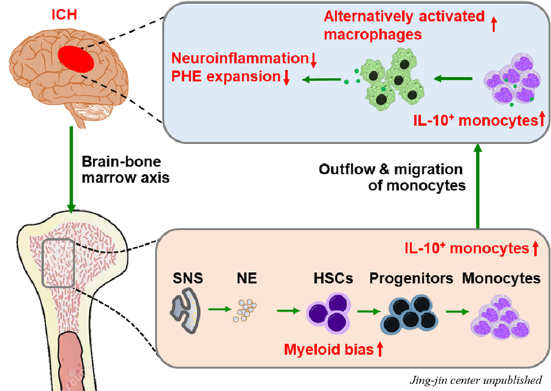
Figure 3: Intracerebral hemorrhage rapidly activates bone marrow hematopoietic stem cells through innervation. Acute period after cerebral hemorrhage, the body activates the bone marrow hematopoietic system through sympathetic nerve innervation. The rapid proliferation of hematopoietic stem cells and downstream myeloid progenitor cells leads to the formation of a large number of immunosuppressive monocytes in the bone marrow in a short period of time. These monocytes then migrate to areas of brain damage. Through neuromodulation or immune intervention, more mononuclear cells with immunosuppressive effect can be generated in the bone marrow after intracerebral hemorrhage, so as to suppress neuroinflammation after intracerebral hemorrhage and reduce cerebral edema.
Astronomical and cerebral imaging: applications of neuroimaging and visualization techniques
General and molecular imaging techniques such as OCT and MRI can also be used to observe the structure and function of the brain. The TANGO trial, published in The Lancet Neurology in May, used The two techniques to track changes in The eyes, brain and spinal cord of patients with Neuromyelitis Optica Spectrum Disorder (NMOSD) during treatment (Figure 4). TANGO trial is the result of a large randomized controlled clinical trial led by Professor Shi and coordinated by Dr. Zhang Chao as the first author with six hospitals in China. This trial reported that Tocilizumab, an IL-6R blocker, had a significant advantage over Azathioprine, an immunosuppressive agent, in reducing the recurrence and disability rates in PATIENTS with NMOSD, providing advanced (grade I) evidence-based medicine evidence for the treatment of the disease. The TANGO trial is a randomized, controlled, investigator-designed, self-funded, independent of any pharmaceutical company, and the world's first comparative, head-to-head study. The Results of the TANGO trial will greatly facilitate the entry of targeted IL-6R therapies into the international guidelines for NMOSD, which will guide neurologists worldwide on how to select new targeted therapies and traditional immunosuppressants.
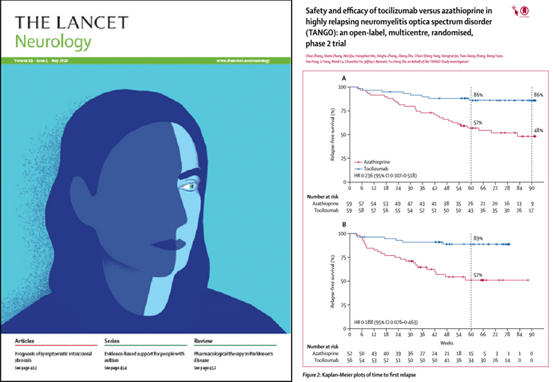
Figure 4: Left: The main result of The TANGO trial was published in The Lancet Neurology (May 2020). Right: Tozumab significantly reduced the risk of recurrence in patients with neuromyelitis spectrum disease compared to azathioprine.
The team recently used single-cell sequencing technology to systematically evaluate the development, differentiation, and autoimmunity antibody production of B cells in bone marrow, blood, and cerebrospinal fluid in NMOSD patients, revealing the molecular mechanisms of peripheral and central activation of autoreactive B cells, as well as immune tolerance loss. It brings new light to the development of new therapies for NMOSD precise immune intervention (Figure 5).
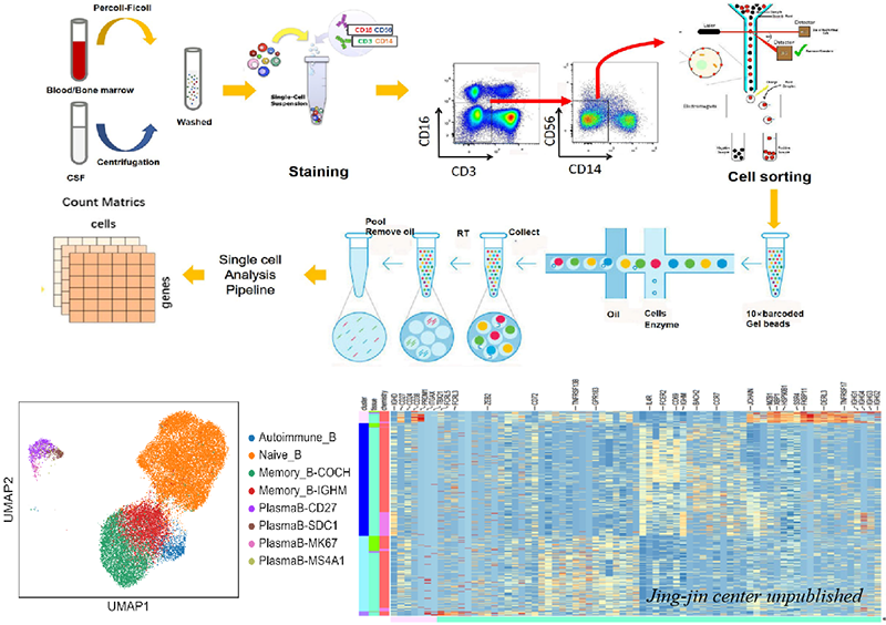
Figure 5: Single cell sequencing experiment flow of B cells in NMOSD patients. NMOSD patients peripheral blood, bone marrow and cerebrospinal fluid of tissue samples, using flow cytometry sorting to obtain high purity of B cells, and then in 10 ´ Genomics single-celled sequencing platforms to transcriptome sequencing of B cells, realize the rapid and efficient single tens of thousands of cell markers, sequencing and analysis, to obtain a single B cell level of the gene expression patterns and differences. Based on each group of B cell division and cell differentially expressed genes between detection, and the analysis of the large-scale, efficient processing tens of thousands of B cells and different subgroup of transcription reaction, so as to reveal the memory B cells and autoimmune B cell activation and amplification and cloning of reactive antibody molecules.
Ultra high field MAGNETIC resonance imaging (UFMR) at 7T enables submillimeter level 3d high resolution imaging, thus revealing tissue details and minor lesions that are difficult to present on 3T. The neural activity contrast of the functional imaging on the ultra-high field platform was significantly enhanced, and the spectrum width expansion of the spectrum scanning promoted the high-resolution resolution of different components. This provides a powerful new tool for peering into the mysteries of brain disease (Figure 6).
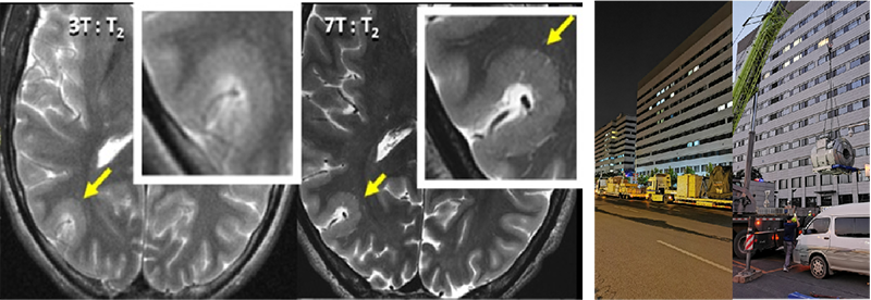
Figure 6 left: Contrast of head image between 3T-MRI and 7T-MRI. Right: National Neurological Disease Clinical Research Center / 7T MRI machine of Beijing Tiantan Hospital.
With the development of brain visualization technology, the team, in cooperation with Academician Cheng Heping's team in Nanjing Brain Observatory, used a miniature two-photon microscope to observe for the first time the abnormal discharge of neurons during epileptic seizure in awake mice, that is, the specific electrical activity of neurons in synchrony, high-frequency and peak disorder when convulsion occurs. This work, using cutting-edge technology, promises to shed light on new mechanisms for the development of epilepsy (Figure 7).

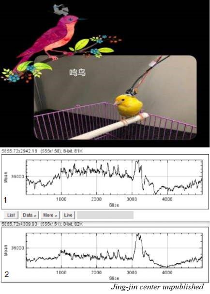
FIG. 7, left: Representative images of neuron-specific electrical activity in M1 region (1-9 represents 9 randomly selected neurons and 10 represents background fluorescence intensity) in convulsive mice. Bottom right: Quantitative analysis of the electrical activity of corresponding neurons. The amplitude of fluorescence change (△F/F0) showed the electrical activity of neurons in the specified time window in epileptic state.
Brain fluid biopsy: fluid markers
The proteins, DMAPS, and immune responses produced by various neuropathic injuries and degenerative changes can reflect chronic, subclinical, subtle changes that are usually not detectable by current imaging methods. These signals can be detected in cerebrospinal fluid, peripheral blood, and dynamically reflect brain changes while overcoming the cost and damage associated with repetitive MRI, PET, and brain biopsies, known as "fluid biopsies" or humoral markers of the brain. These methods provide irreplaceable means to track the progress of brain diseases and thus can be used as brain fluid Windows. Neurofilament protein (NfL) has been widely recognized as a marker of multiple sclerosis progression. The value of the NfL in predicting neurodegeneration and other diseases is being revealed (Figure 8). Chen Jingshan, Zheng Pei et al. first detected the expression level of neurofilament protein (NfL) in healthy Chinese population, and established a reference for the NEGATIVE interval of NfL for Chinese people. A positive correlation was found between NfL plasma levels and age in healthy individuals, and it is suggested that the NfL negative interval should be divided by age. Further in stroke patients, it was found that NfL's prediction of stroke progression and prognosis was significantly better than other biomarkers. For unknown new humoral markers, Shi introduced the team's work of screening humoral markers based on proteomics. The proteins isolated from the patient's plasma or exosomes were identified by mass spectrometry proteomics. After the differential proteins were screened, their expression was verified by ELISA or hypersensitivity detection platform according to the different kurtosis of the proteins. Combined with imaging and clinical data, a combination of humoral markers for differential diagnosis, curative effect and prognosis assessment was selected (Figure 8). As for the next step in the research and development of body fluid markers, combined with multiple omics and artificial intelligence methods, combination screening of inflammatory related markers for neurological diseases will be carried out, so as to find humoral markers and drug intervention targets for auxiliary diagnosis and prognosis assessment, and further explore the pathological mechanism by using animal disease models.
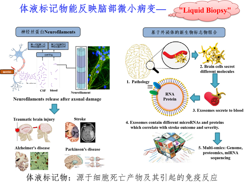
Figure 8. Left: AFTER axonal injury caused by nervous system disease, NfL is released into the cerebrospinal fluid and enters the peripheral blood through the blood-brain barrier. Right: After the neurological damage caused by pathological changes in the brain, cell damage-related molecules are released into the peripheral blood through exosomes. Proteins and RNA contained in exosomes are related to the severity of the disease, which can be screened and identified by multiple omics techniques (including proteomics and transcriptomics).
In recent years, with the continuous update of laboratory testing methods and means, a number of domestic units or companies have established related tests for neuro-immune diseases based on their own conditions. However, there are still many differences in the testing methods and systems used by different centers, which lack unified industry standards. Moreover, the testing kits approved by CFDA are very limited at present. For the detection of autoantibodies expressed by neuroimmune disease membrane, for example, the most specific assay was cell-based indirect immunofluorescence (CBA) assay, which was widely recognized for its ability to maintain the self-conformation and protein modification of antigens. However, there was no CFDA approved CBA assay kit at present. Based on this situation, Professor Shi described the team's years of commitment to the establishment of a laboratory diagnostic platform for neuroimmune diseases and technological innovation, and the team's support for the formation of the R&D transformation team - Tianhai New Domain. At present, Professor Shi's transformation team has established a mature neuroimmune disease detection system and a new humoral marker development platform, including hypersensitive peripheral blood detection platforms based on cellular immunity, western blot, immunomagnetic particle detection, and single molecule, as well as a new humoral marker development platform based on multi-omics. The aim is to create independent intellectual property rights, world-class technology and quality control for promotion in China and internationally.
New Terrain: www.newterrain.cn Lab for life

Figure 9 market transformation team of humoral diagnosis of neuro-immune diseases -- New Terrain
Brain networks and hospital information networks: Trickle into sea
Whole brain mesoscopic neural connections map of micro optical sectioning tomography imaging informatics and able to sketch a brain space neural connection graph, pathological model under specific neural circuits for fine mapping, to research and development in the early diagnosis of major diseases, brain precision intervention (including disease before intervention and treatment after rehabilitation) of the new technology, new method is of vital importance (Li, et al. Science 2010; Gong et al. Nature Communications 2016). Qing-ming luo academician brain space informatics (Brainsmatics) concept is put forward, aimed at the brain as a complete system, comply with the demand of intelligent technology development, with the basic theory of brain science, system science and information science as a guide, using the emerging whole brain high resolution accurate spatial positioning and imaging method, combined with a variety of advanced technology to study the brain at the same time, tag, acquisition, analysis, and visualization have clear space scale and the location of the fine brain network structure and function information, extracted from large data across level, brain connections, the space-time characteristics of the multi-scale perception, emotion and consciousness such as brain connection mechanism of spatial information, Thus promote the leapfrog development of brain health and intelligent technology.
The National Hospital Quality Monitoring System collects the full coverage and diagnostic information of the national public hospital (HQMS), similar to the brain network and brain information aggregation, analysis and processing (Figure 10). Professor Shi introduced that the team led by Tian Decai used HQMS to calculate the incidence rates of multiple sclerosis (MS) and optic neuromyelitis spectrum diseases in all age groups and 31 provinces and cities in China, both of which were published in Lancet Regional Health (Figure 10). This study filled in the gaps in the epidemiological data of MS and NMOSD of nearly 1.4 billion Chinese people, improved the map of the incidence of MS and NMOSD globally, and provided a basis for the formulation of national health insurance policies.

Figure 10 left: Professor Shi introduces the National Platform for Information on Neurological Diseases, similar to the brain gathering and processing information, to the UCB Asia Pacific Director. The platform implements dynamic monitoring of the diagnosis and treatment of neurological diseases in 31 provinces, autonomous regions and municipalities directly under the Central Government. Right: MS and NMOSD are mainly involved in young women by using the traditional Image of female flower women in China. Although MS and NMOSD are sick and thin, they have bright eyes and full of expectation for the cure of the disease. Background: This work was completed at the National Center for Clinical Research of Neurological Diseases, based in Beijing Tiantan Hospital.
Window of the brain: God meets the sky
Professor Shi's report combines his curiosity and exploration of the brain as a neurologist with his passion. By uncovering the mysterious veil of the brain, he aims to reduce the pain of brain diseases, stimulate the conscience and potential of human beings, and overcome the common difficulties faced by human beings. He is full of optimism and yearning. His closing words: If you stand on the summit, you will gain a magical view of the sky and the earth; If you can join us in harnessing the latest in science and technology, in finding the window of the brain to defeat brain diseases, in tapping into the human intelligence that we are currently facing in an effort to overcome the unprecedented plight of humans and all species, you will see our beautiful mind.